
Modality
Magnetic Resonance Imaging
Introduction
Magnetic resonance imaging, more commonly known as "MRI", is a sophisticated method of imaging the body which, unlike CT and radiography, does not use ionizing radiation. MRI capitalizes on the molecular composition of tissues to produce an image.
Unlike with CT, images can be obtained directly in the axial, coronal, or sagittal planes, and 3 dimensional volumes of data can also be obtained. Advantages of MRI include the excellent soft tissue contrast and differentiation, reducing the need for intravenous contrast enhancement, the ability to evaluate blood flow, and the ability to subtract out background tissues that are not of interest.
The goal of this module is to serve as an introduction to MRI, and to provide you with information to aid in understanding what you are looking at when you view an image.
You can view individual sections of this module using the selections on the right.
How Images Are Obtained

MRI is a technically complex imaging modality, and it is beyond the intentions of this module to discuss image acquisition in great detail. The following provides a brief summary of how MRI images are obtained and viewed.
The patient lies on a table which is moved into the bore of a large, powerful magnet (most of those in clinical use are 1.5 Tesla). Protons in the patient's tissues will naturally align themselves with this powerful external magnetic field.
Radio frequency pulses are sent into the patient, causing disruption of the alignment of the protons (the protons absorb energy and are displaced from their aligned position). Over time, the protons will naturally realign themselves with the external magnetic field. When this occurs, they emit the absorbed energy as radio frequency waves which are detected by the MRI scanner. These emitted radio frequency waves are the MRI signal.
The location and distribution of the emitted MRI signal is detected and analyzed by a computer, and used to produce an image. Thus, a radio frequency pulse is sent in, the patient emits a signal back which the machine reads and uses to create an image.
What types of tissue emit the MRI signal?
Only molecules with an odd number of protons have their own magnetic field and align themselves with the external magnetic field. It is these molecules that are responsible for emitting the MRI signal which is used to create the image.
Hydrogen atoms are the most abundant of these molecules in the human body and provide the basis for MRI imaging. The body is largely composed of water, and the hydrogen atoms in water are responsible for producing much of the MRI signal.
MRI images are displayed in shades of gray, like radiographs and CTs. The shade of gray that a given structure appears on MRI will vary with the MRI technique used to obtain the image. This will be discussed later in this module.

Images can be obtained directly in the axial, coronal, or sagittal plane. Like CT, axial images are viewed as though looking at the patient from the foot towards the head, placing the right side of the patient on the left of the image. Like radiographs, coronal images are viewed as though looking at the front of the patient, placing the right side of the patient on the left of the image. Sagittal images are viewed such that anterior is on the left of the image and posterior on the right.


Factors Affecting the Appearance
A myriad of factors affect the appearance of the final MRI image, including tissue composition, machine parameters such as the timing with which the radio frequency (RF) pulse is sent in and when the machine "listens" for the returning MRI signal, the use of intravenous contrast, and whether the structure being imaged is static or dynamic (i.e. flowing blood).
A complete discussion of this is beyond the goals of this introductory module. It is not necessary to memorize the physics or the appearance of certain tissues on MRI, however you should be familiar with the concepts and realize that the same structures might look different on different types of MRI images.
Tissue Composition

As discussed in "how images are obtained", hydrogen atoms provide the basis for MRI imaging of the body. Tissues which are abundant in hydrogen atoms, such as water and fat, are responsible for producing most of the MR signal. Likewise, tissues which are devoid of hydrogen atoms, such as cortical bone, do not give off much signal.
Tissues which emit a strong MRI signal will appear white on MRI images and are termed "high signal intensity" and those which emit a weak MRI signal appear blacker and are termed "low signal intensity".
This is a sagittal MRI of the cervical spine. Note the cerebrospinal fluid which has a very high water content, has a high signal intensity on this image, appearing white. In contrast, the cortical bone appears black, "low signal intensity" due to its low water content.
Contrast Enhancement
Similar to the use of intravenous contrast in CT, the conspicuity of and contrast between structures of similar composition can be augmented by the use of intravenous contrast in MRI.
The contrast used in MRI is made of gadolinium, a paramagnetic substance with a strong magnetic field. Gadolinium affects the appearance of tissues on T1 weighted images, causing tissues which take it up to appear brighter on T1 weighted images. Gadolinium is administered to the patient by intravenous injection. Tissues that appear brighter on images obtained after gadolinium are said to "enhance".

The first image is a T1 weighted axial image of the abdomen. The next image was obtained after intravenous gadolinium was administered. Note that the kidneys, aorta and pancreas are brighter due to the gadolinium. In addition, the renal medulla and cortex can be differentiated on the enhanced images, as they have taken up different amounts of gadolinium. Tumors and other abnormal tissues take up gadolinium differently from normal tissues, and gadolinium is to augment detection of these.
Unlike CT, not all MRI images are obtained the same way, and as a result, the images can look quite different. MRI machine parameters, such as the strength of the RF pulse, the timing of repetitive RF pulses, the time the machine "listens" for the returning signal from the patient can all be selected and manipulated to create specific types of images and to optimize visualization of certain structures.
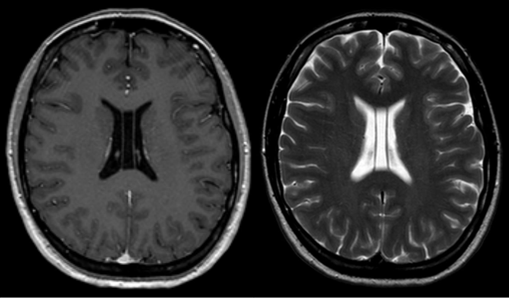
Two common "sequences" or types of MRI images which you should be familiar with are T1 and T2 weighted images. The images are "weighted" to bring out various characteristics of tissues. Some tissues look the same on both T1 and T2 images and some look different.


These sagittal lumbar spine MRI images are both T1 weighted and T2 weighted. Note that subcutaneous fat is bright on both images. The cerebrospinal fluid is dark on T1 and bright on T2. Note the difference in the appearance of the intervertebral discs on the two images. The central portion of a disc contains fluid and so appears brighter on T2 than T1.
It is not necessary to understand the physics or memorize the appearances of tissues on T1 or T2 weighted images. The goal of this section is for you to become familiar with the concepts and realize why the same structures might look so different on different MRI images or "sequences".
The two images the below are the same axial section of the upper abdomen. The image on the top is T1 weighted and the image on the bottom is T2 weighted. Note the fluid filled gallbladder and the cerebrospinal fluid are different shades of grayscale on the two types of images. Also note that the soft tissues, such as the liver, appear slightly different shades of gray on the two sequences.

Blood vessels, such as the aorta (labeled above) are even more complex. Not only do the machine parameters affect their appearance, but the speed and direction in which the blood flows also impacts the final image.
Anatomical Structures
MRI provides excellent soft tissue contrast and can readily demonstrate and differentiate adjacent soft tissue structures, far superior to that possible with CT.
Everything is so beautifully seen on MRI that it is easier to discuss what isn't well seen. Air does not give off an MRI signal and produces a void on MRI, thus air containing structures such as the lung appear black. Likewise, MRI of the bowel is limited due to the presence of intestinal gas. Bowel peristalsis also causes motion artifact, limiting the utility of MRI in the evaluation of the bowel.
Abdomen
The images below were both obtained without iv contrast enhancement. The image on the top is a CT and the image on the bottom is a T2 weighted MRI. Note that on the CT all of the blood vessels and solid organs (liver, pancreas, spleen and kidneys) appear the same shade of gray. Note that on the MRI all of these structures are subtly different shades of gray, allowing us to distinguish them. Also note that the portal vein can be discretely seen from the adjacent pancreas on MRI, and individual nerve roots of the spinal canal are visible.


The image below is a T2 coronal MRI image of the abdomen. Note that the lungs appear black, since air does not give off an MRI signal.

The next image is a T1 axial image of the abdomen. Note the fuzzy, ill defined margins of the bowel. This is caused by motion artifact, as the bowel is constantly moving (peristalsing). In the evaluation of bowel, CT is superior to MRI. The remaining soft tissues are well demonstrated, including the muscles of the back and abdominal wall.

Extremities
Ligaments, tendons and cartilage are easily displayed and evaluated on MRI, as demonstrated on the below coronal MRI of the knee. This "internal anatomy" of joints can only be seen with MRI, not with any other type of radiologic imaging.
Soft tissues of the extremities are also better evaluated with MRI than other imaging modalities, as demonstrated on the next image which is an axial MRI of the thigh. The medium gray muscles can be differentiated from the darker tendons, and the bright intervening fat helps to make individual muscles conspicuous. Note that the sciatic nerve is even discretely seen!

Important to note is that dense cortical bone, such as of the femur, appears black. Cortical bone has a low water content and gives off very little signal. CT is superior for evaluating cortical bone. In contrast, the medullary bone, containing the bone marrow, does give off MRI signal and can be adequately seen on MRI.
Pelvis
These are axial and sagittal T2 weighted MRI images of the female pelvis. The uterus and ovaries are beautifully displayed on MRI. The different soft tissue components of the uterus can be differentiated on the sagittal image; the endometrium is bright and the myometrium is darker. Note that the ovarian follicles are seen as white (fluid filled) structures. The final image shows CT image of the pelvis to compare with the MRI images. The rectum, pelvic sidewalls, prostate and seminal vesicles are also well seen on MRI.

Brain and Spinal Column
The following images show an axial T2 MRI of the spine, and an axial T2 MRI of the brain which includes the orbits.
The darker spinal cord is seen discretely, surrounded by the brighter subarachnoid fluid. The white dot in the center of the spinal cord is an abnormal fluid filled cavity, called a syrinx. The nucleus pulposis, the fluid component of the intervertebral disc, can be differentiated from the annulus fibrosis, the outer dark fibrous band of the intervertebral disc.

These images demonstrate how exquisitely the internal anatomy of the brain and spinal cord is seen. No other imaging modality can compare with MRI for imaging the central nervous system.
Indications and Specialized MRI Techniques
Since MRI has excellent soft tissue contrast, its greatest use is for evaluating soft tissue structures, as discussed previously in this module. MRI is indicated in the evaluation of joints and soft tissue tumors and masses of the extremities. In the neck, chest, abdomen and pelvis, MRI is indicated in the evaluation of congenital or structural abnormalities, tumors or other masses of organs and soft tissues. MRI is indicated to evaluate the brain, face, spinal cord and spine for a wide variety of symptoms and problems. Specialized techniques include specifically imaging blood vessels and fluid filled structures.
Tumors
MRI is widely used to evaluate for tumors throughout the body, including the neck, chest, abdomen, pelvis, extremities, spine and brain. It is excellent in the evaluation of brain tumors. The two images demonstrate brain tumors in two different patients. The images were obtained after IV contrast was given, and the tumor tissue has taken up the contrast and appears brighter than the adjacent brain parenchyma on these T1 weighted images.


Magnetic Resonance Angiography (MRA)
This specialized technique takes advantage of the flow properties of blood vessels to obtain images. MRA is commonly used to evaluate arterial blood flow to the central nervous system. It is also used to evaluate arterial blood flow to the extremities, abdominal organs and the intestines.
MRA displays the caliber of a blood vessel, allowing evaluation for occlusions, stenosis or aneurysms, for congenital abnormalities, and for the presence of blood clots or atherosclerosis.
The image below is an MRA of the arch of the aorta, the subclavian arteries and arteries of the neck. Note the arteries appear bright, while the background tissues are dark and not well seen with this specialized angiographic technique.

Magnetic Resonance Venography (MRV)
This specialized technique takes advantage of the flow properties of veins to obtain images.
MRV is commonly used to evaluate venous drainage from the central nervous system. The image below is a sagittal view of an MRV of the brain. Flowing blood in the veins appears white while the background tissue is poorly seen. Blood flow in the arteries is not seen with this technique.

MRV is also commonly used to evaluate the veins of the chest, abdomen, pelvis and extremities, to detect abnormal areas of narrowing or dilatation or for the presence of blood clots.
MRCP
Magnetic Resonance Cholangiopancreatography
This specialized technique takes advantage of how bright fluid appears on T2 weighted images in order to specifically display fluid containing structures. The bile within the biliary ducts of the liver, the common bile duct and gallbladder, and the fluid in the pancreatic duct all appear very bright on these specialized images, while the background tissue is poorly seen.

MRCP is commonly used to evaluate for obstruction of the bile ducts or pancreatic ducts, for stones within the ducts or gallbladder, and for congenital anomalies of these structures.
A normal coronal MRCP is shown above. Note how well the fluid containing structures are seen. The stomach and duodenum contain fluid in this patient, so they are visible as well.
Quiz
Using the T2 weighted image on the right for guidance, what shade of gray would you expect the following tissues to look like on T2 weighted MRI?

this is correct
what about this one