
Modality
Radiography
Introduction
Radiographs, which are commonly called X-rays, are two-dimensional images of the body that are displayed as white, black, or varying shades of gray. Radiographs are utilized throughout all specialties of clinical medicine, and they are widely accessible and fairly inexpensive.
This module serves as an introduction to radiography. It discusses basic principles and will supply you with information to help you understand what you are looking at when you view an image. What causes a structure to look black, white, or gray on a radiograph? Why are some structures seen better than others?
How Images Are Obtained
Radiographs are images that are obtained using ionizing radiation. An X-ray passes through a patient to hit a specialized detector. Historically, images were created and printed on film. Currently, radiography has become digitalized and the images are viewed on specialized computer workstations.
X-rays are discrete quantities of electromagnetic radiation that are produced outside the nucleus of an atom. X-rays are produced in a generator by the interaction of an electron beam with a Tungsten target. The X-rays are emitted from the generator as a beam and travel through a patient. Tissues absorb (attenuate) some of the x-rays, while others pass through and hit the cassette. The attenuation properties vary among different tissue types, resulting in a heterogeneous distribution of X-rays emerging from the patient to hit the detector (historically called a cassette).

Screen-film radiography: The cassette contains film and intensifying screens. When the X-rays hit the film, a photochemical interaction occurs, causing the metallic silver in the film to precipitate. This renders the film black when it is developed chemically.
Digital (computed) radiography: The cassette contains a photo-stimulable phosphor detector system. When the X-rays hit the cassette, a fraction of the absorbed energy is trapped in the detector. A red laser light stimulates the emission of the trapped energy, and blue-green light is emitted, collected, and converted into an electric signal. This is digitized and stored as a pixel value, with the gray scale dependent upon the amount of energy absorbed & released.
Viewing the Image

A radiograph is a 2 dimensional image. It is a summation of all of the structures through which the x-ray beam passed as it traveled through the patient. Structures are superimposed on the film, and depth cannot be determined; when viewing an image, you cannot tell if a structure is anterior or posterior.
Films are viewed as though you were standing face to face with the patient. The patient's right side is displayed on the left of the image, and the left side on the right.
The more that the x-ray beam is absorbed by the tissues it passes through, the lighter the structure appears on the X-ray image. Tissues that absorb more of the X-rays look whiter on the image, such as the ribs, while tissues which absorb less of the X-rays appear blacker on the image, such as air.
Factors Affecting the Appearance
The composition, thickness and shape of a structure all interact to influence what an object looks like on a radiograph.
Density
What causes a structure to appear black, white, or gray on a radiograph? It is dependent on how much of the x-ray beam is absorbed by the structure vs how much passes through to reach the detector. The density of the object being imaged determines how much of the x-ray beam will be absorbed.
Dense structures absorb (attenuate) more of the x-ray beam than less dense structures. Thus, less of the beam passes through to hit the cassette and these structures appear white, termed radioopaque. The radiograph shown is a pair of metal scissors, which are radioopaque. Note the scissors are brighter white in areas where the metal is thicker, like the handles. Other dense structures include calcium, barium and iodine, all of which look white on radiographs. Barium and iodine will be discussed in later sections of this module.

Structures which are not very dense, such as air, absorb very little of the x-ray beam. Most of the beam passes through the air and strikes the detector. As a result, these structures appear black on x-rays, termed radiolucent. Note that on the radiograph shown, air surrounding the scissors is black.
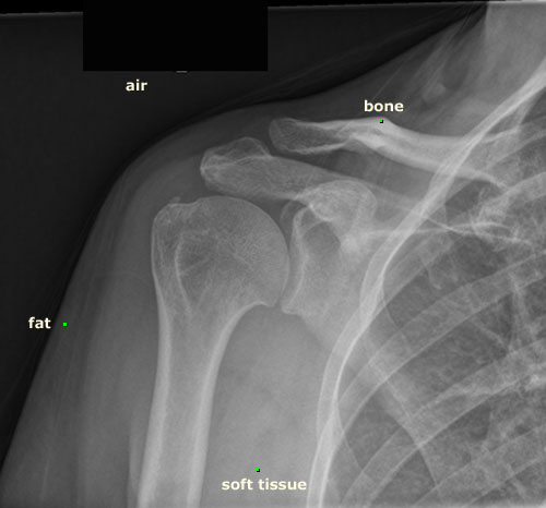
The variable densities of structures in the body result in the four basic radiographic densities:
- Air - black
- Fat - gray/black
- Soft tissues and organs - gray
- Metal, calcium, and bone - white
Fat is low density, but is slightly greater than air and so looks less black on a radiograph than air. Muscles, organs, and soft tissues are shades of gray, ranging from light to dark gray depending on the structure’s density. These shades of gray are referred to as water density.
This radiograph shows three cups containing substances of different densities. Which is the most dense?

The cup on the far right is empty, containing only air, which is very low in density and therefore appears black. Low density structures are also called radiolucent.
The center cup contains water. Fluid is denser than air, and thus it will attenuate more of the beam and appear an intermediate shade of gray.
Do you know what is in the cup on the left? It is barium, a very dense liquid which attenuates much of the beam and so appears white on radiographs. For some radiology procedures, patients drink barium to opacify the gastrointestinal tract and make it visible using x-rays.

Thickness

The thicker a structure is, the more of the x-ray beam it will absorb (attenuate). As a result, thicker structures will appear whiter on radiographs than thinner structures of the same composition.
The top two images on the right are photographs of a metal step plate, one from the side view (lateral) and the other from above. The plate is made of a uniform material of successive thickness; section 1 is the thinnest and 10 the thickest. The lowest image on the right is a radiograph of the same step plate taken from above. Note that the thicker segments of the step plate are more radiodense. The variations in the grayscale correspond to changes in plate thickness.
Below is a radiograph of two cups of water. Notice that they are the same shade of gray on this image because the thickness (diameter) of each cup is the same.

How do you think each would look if a radiograph was taken of them from the top (looking down into each glass)? Why might they appear different shades of gray when imaged from above?
This is a radiograph of the same cups of water taken from above. Why does the cup on the left look whiter?

Remember that the cup on the left contained more fluid than that on the right. Therefore, more of the x-ray beam was attenuated as it passed through, causing a whiter appearance on x-ray.
Shape
The shape of an object will affect its final appearance on radiography. The shape affects how much of the x-ray beam is attenuated by a structure, mostly because of differences in thickness. For example, if looking at a round structure of uniform composition, like a solid ball, the center will appear denser, or whiter on a radiograph than the edges because the center is thicker and attenuates more of the x-ray beam.
The radiograph below shows a more complex structure; a green pepper.

As you know, the inside of a pepper is comprised of air, and the walls are the "flesh". Area B appears more lucent (black) than area A, despite the fact that the outer wall of the pepper is of uniform thickness. This is because the x-rays passing through area B had to go through the flesh of only the front and back wall, while those passing through area A had to pass through much more of the flesh, as the beam traveled down the barrel where the wall of the pepper curves. As a result, more of the beam was attenuated and the lateral walls of the pepper appear whiter.


Do you know what these radiographs are of? That on the left is taken from above and that on the right from the side. It is an artichoke, and as you can see, has a complex appearance on radiography. Note that the outermost leaves, which can be seen in isolation, are very lucent, except at the tips where they curl in somewhat and appear denser due to some overlap. The choke, or center of the artichoke is the whitest area, due to its density and thickness in comparison to the fine leaves. The image taken from the side shows the air intervening between the leaves.
What Anatomical Structures Are Well Seen
Radiodense structures are easy to evaluate on radiography. Examples of these include bone, calcium, barium and metal. Radiolucent structures are also easy to evaluate. Examples of these include air and fat. Structures of intermediate density, such as fluid and soft tissues like organs and muscles are harder to evaluate and differentiate. They all appear a similar shade of gray. How distinctly they are seen on radiographs depends on what structures they are next to.
If two structures of similar density are adjacent to one another, they cannot be differentiated since they appear the same shade of gray. In contrast, adjacent structures of differing densities, like bone and fat, can be distinguished quite easily.

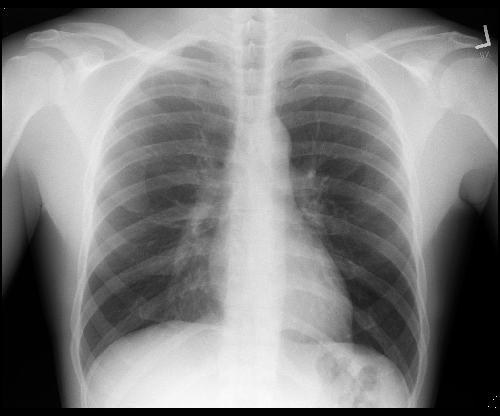
Let's use this radiograph to examine how various tissues look on x-ray. Bones are radiodense and appear white. Look at the ribs and clavicles.
Air does not attenuate much of the x-ray beam. Structures that don't attenuate much of the beam appear black ("radiolucent"). Since the lungs are composed primarily of air, they appear black on radiographs.
Soft tissues are displayed in varying shades of gray. They are only well seen when there is tissue next to them of very different composition. For example, on the CXR, the edge of the heart is seen discretely from the lungs due to the significant difference in their densities. However, one cannot differentiate internal structures of the heart or mediastinum (like the aorta and pulmonary arteries) because these tissues have similar densities, attenuate the X-ray beam similarly, and therefore look the same on x-rays.

This is a radiograph of the upper abdomen.
Most of the abdomen is occupied by soft tissue structures which are difficult to differentiate from one another. The gas that fills the stomach and bowel (intestines) appears black and makes these structures visible. The bowel that is filled with food or fluid is not well seen, as it blends into the background gray appearance of the other soft tissue structures of the abdomen.
Fat is slightly more radiolucent than other soft tissues and appears blacker; see the preperitoneal fat stripe in the right side of the abdomen.
Indications For Use
Radiographs are commonly obtained to work up a wide variety of problems and symptoms of the chest, abdomen, pelvis, extremities and spine. Radiographs are relatively inexpensive, widely available, and easily accessible.
It is beyond the intentions of this submodule to discuss all of the potential uses of radiography, but this should serve as a foundation to help one understand some of the general uses.
Bones
As bones are easily visible on radiographs, X-rays are a great way to evaluate for bone pathology. The outer edge of long bones, called the cortex, is more dense than the inner portion, the medulla. Disruptions of the cortex, as occurs in fractures, are readily seen on x-ray.
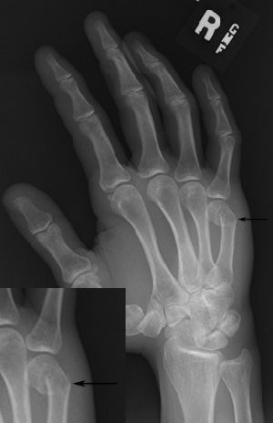
This hand radiograph reveals an acute fracture of the 5th metacarpal (arrow). The smooth outer edge of the bone is disrupted at the site of the break. Note the bone distal to the break is angulated anteriorly. This is known as a "boxers" fracture, as the mechanism of injury is usually a punch, where the force is transmitted to the 5th metacarpal bone. Below is a magnified view of this fracture.

This radiograph of the right shoulder shows an acute fracture in a child.
The black arrows point to the fracture line. On the medial side, it is easy to see the disruption of the medial cortex of the bone.
The fracture line itself is more lucent than the adjacent bone, with the space between the bone fragments filled with blood and marrow, which are less dense than bone.
The clue that this is a child is the growth plate, which is a smooth, undulating lucent line separating the epiphysis from the metaphysis.
Disturbances in bone growth, including premature fusion of the growth plate, or abnormal widening of it, can be detected with radiographs. X-ray evaluation of the bony structures and growth plates of the hand are used to determine bone age.
In addition to fractures, radiography is an excellent way to look for focal lesions like tumors or infections that cause bone destruction. Other indications for use include evaluation of joint spaces for infection, arthritis, and dislocations.
Chest
Chest radiographs are one of the most common radiologic studies obtained.
They are excellent for evaluating the lungs, as normal lung appears black and most abnormalities, like infections, fluid collections and tumors, will appear varying shades of gray.
Can you see the lung mass on this CXR?

Masses are densely packed soft tissue, so they can be differentiated from the adjacent lucent normal lung quite well. Look below to see if you correctly identified the mass.

Chest radiography is also used to evaluate the size and shape of the heart and other mediastinal structures, and for detecting abnormalities of the pleura, such as a pleural effusion. A pleural effusion is an accumulation of fluid within the pleural space, between the parietal and visceral pleura. The fluid may be of a variety of compositions, including blood, pus and more simple fluid.
The radiograph shows a right pleural effusion. Fluid is much denser than air, and the contrast between the normally aerated, radiolucent lung and the denser fluid is sharp. Most of the right side of the chest is filled with fluid. A meniscus is seen along the lateral chest wall, as indicated by the arrow.

Abdomen
Radiography is commonly used to evaluate the intestines (small and large bowel). The stomach and intestines contain both air and fluid. The radiolucent air makes the intestines visible on radiography.
This radiograph shows a small amount of air in the small bowel, and a bit more air is visible within the colon and rectum. If the intestines are abnormally dilated, as occurs in a bowel obstruction, this is easily identified on a radiograph. Kidney stones, which contain calcium, are also readily evident on abdominal radiography as dense foci.

It is more difficult to identify the solid organs of the abdomen and pelvis, since they are all of similar soft tissue density. The edges of these organs can be seen only if they are separated by tissue of differing composition, for example fat.

In the radiograph above, the inferior edge of the liver and kidney are visible because of the contrast with the fat adjacent to them, which is more radiolucent and looks blacker on the film. In this patient, there is also a small amount of air in the stomach. Try to find these areas on the radiograph above and then check you answers below.

Dense structures, such as calcifications and metal, are easily seen on radiographs. This patient swallowed a pin, which can be faintly seen overlying the sacrum in the midline.

