Modality
Ultrasonography

Introduction
Ultrasonography uses high frequency sound waves to create images. The sound waves are directed into the body and are reflected back to the ultrasound transducer. The reflected sound waves provide information about the depth, type of tissue, and tissue interfaces. This information is converted into grayscale images.
Unlike radiography, fluoroscopy and CT, ultrasound does not use ionizing radiation, so is low risk to the patient. It is non-invasive, relatively inexpensive, and allows one to perform "real-time" or "live" imaging and to observe motion.
How Images Are Obtained
An ultrasound transducer (shown below) is placed onto the patients skin with the gray area in full contact with the skin. Gel is used between the skin and transducer to prevent air from intervening between the two. The transducer emits a narrow beam of sound waves of a specific high frequency. Depending on the frequency, the sound waves penetrate the patient's tissues to a variable depth and are refracted, scattered, absorbed or reflected. The reflected waves (called an echo) bounce from tissues back to the transducer, which also acts as a sensor, or detector, and are used to generate the image.

High frequency sound waves provide high resolution images, but don't penetrate deeply into tissues, so are best for superficial structures. Lower frequency sound waves are used to image deeper structures, but create lower resolution images.
Sound waves are reflected at the interfaces of tissues with different acoustic impedance (a product of the density of the tissue and the velocity of sound in the medium). The intensity of the reflected US beam depends upon the different characteristics of adjacent tissues and tissue interfaces. Larger differences in tissue types yields higher intensity of the reflected sound. High intensity echos appear white on the US image, "echogenic or hyperechoic". Low intensity echos appear dark, or "hypoechoic".
An US scanner is shown below. As the transducer is moved over the patient, the screen displays the image in real-time. Since the transducer can be held in any position, images in the axial, sagittal, coronal planes, as well as any obliquity can be generated.

Factors Affecting the Appearance
Composition of the Tissue
Solid organs, like the liver, have many internal tissue interfaces such as the bile ducts, portal vein branches, hepatic artery branches, hepatic veins, peri-portal fat, etc. This results in multiple reflected echos of variable intensity, and so the liver looks a medium shade of gray on US. Organs are slightly different from each other in their shade of gray on ultrasound based on differences in their tissue composition.
Fluid filled structures (gallbladder or urinary bladder) have no internal tissue interfaces and therefore do not reflect sound waves. As a result, the fluid filled portion of the structure will appear black and the walls gray to white. The US image of the gallbladder below illustrates this.

Composition of Adjacent Tissues
The greater the differences between two adjacent structures, the greater the reflected sound waves and the more echogenic (white) the border between the tissues appears on the image. The difference in acoustic impedance between bone or air and adjacent tissues is large, and thus at such an interface most of the sound waves are reflected. Very few sound waves are left to penetrate and reach deeper structures. As a result ultrasound can't be used to image the area deep to bone or air, and since no echos are reflected from these locations, they look black on the US image.
The image below is a superficial ultrasound of the chest wall. The ribs, seen in cross section, are marked with stars. Note the anterior edges of the ribs are echogenic, and the black "shadow" posterior to the ribs, where no sound has been transmitted. The lung edge, running just posterior to the ribs, is also echogenic, but the lung parenchyma can't be imaged and appears black

Anatomical Structures
The solid organs of the abdomen, including the liver, kidneys and spleen, can be evlauated well with US. Deeper structures can't be seen with the same level of resolution as superficial structures because of the number of scattered, absorbed and reflected sound waves occuring, leaving less sound available to penetrate deeper tissues and reflect back to the transducer. As an example of this, look at the difference in resolution of the upper vs the lower pole of the right kidney, shown below. The lower pole, on the right side of the image, is more superifical and is seen with better resolution than the upper pole.

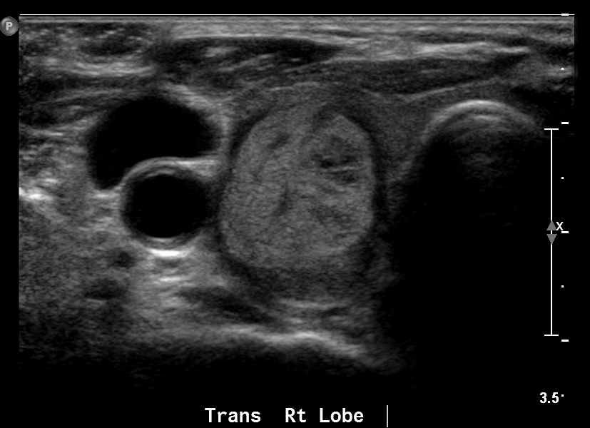
Superficial structures (like the thyroid and testicles) can also be evaluated with ultrasound, with production of high resolution images. The image below is of a testicle. The black (anechoic) structure posterior to it (*) is an abnormal collection of fluid around the testicle, called a hydrocele.

US is widely used in obstetrics (OB), as the fluid in the amniotic sac provides a great "acoustic window" through which to image the fetus. The two images below are an US of a 19 week old fetus. The amniotic fluid is black (anechoic). The top part of the images shows the medium gray of the uterine wall and abdominal wall. Just deep to this is the black amniotic fluid, and then the fetus. The left image is a sagittal image of the fetus. The right image shows the fetus's hand in the center of the image.


Fluid or blood filled structures, for example the gallbladder, urinary bladder, and the heart, can be evaluated well with US. Depending on their location and depending on the composition of adjacent tissues, some blood vessels are seen well. For example, the descending thoracic aorta cannot be seen with ultrasound, due to its location deep to bone and air.
Indications for US
Pregnancy
US is used in early pregnancy to confirm the presence of an embryo. In mid pregnancy, a "screening" ultrasound is performed to evaluate for fetal anomalies. The brain, heart, stomach, kidneys, umbilical cord and other structures are carefully evaluated to determine if they are morphologically normal.
The image on the right shows the fetal head in the axial plane. The outer skull is not yet ossified, which is why we are able to US "through" it. The stars depict the right and left cerebellar hemispheres. The + symbols are located on the thalamus. Identifying these normal structures and confirming their normal size helps to exclude major congenital anomalies.

Gynecology

Beautiful images of the uterus and ovaries are generated when US is performed with a transvaginal transducer (shown on bottom left). It is inserted into the vagina and positioned in the vaginal fornix. This places the transducer right next to the uterus and close to the ovaries, enabling high resolution images to be obtained.
In the image to the right, the vaginal wall is depicted by the green dots. The transducer sits against the wall and you can see its proximity to the uterus. The image on the bottom-right is a normal ovary, with the stars depicting follicles.


Abdominal Organs
The liver, kidneys, and spleen are routinely evaluated by ultrasound. Cysts, tumors, kidney stones and gallbladder calculi are some of the findings which can be seen with ultrasound. The below image on the left is a transverse US of the epigastrium. The body of the pancreas is clearly depicted (marked with the stars), which is unusual because gas within the stomach, lying anterior to the pancreas, often obscures it from being imaged.
It is easy to distinguish blood vessels from parenchyma. The image on the right is a transverse US of the upper liver. This shows the confluence of the right, middle, and left hepatic veins which are marked with stars.


Specialized Technology
Color & Spectral Doppler Ultrasound
Blood vessels can be identified with grayscale ultrasound, as shown above. Doppler ultrasound is used to gain additional information about the blood vessels and the blood flow through them. It is used to measure the velocity and direction of blood flow, to evaluate for stenosis (narrowing) of blood vessels, and for vascular resistance and patency.
Doppler US relies on the doppler effect; the change in frequency of sound waves reflected by a moving object. Blood moving towards the US transducer appears to have a higher frequency and shorter wavelength, while that moving away from the transducer appears to have a lower frequency and longer wavelength.
The image on the left is a color (power) doppler image showing blood flow in the common, internal and external carotid arteries. The image on the right is a spectral doppler image of the internal carotid artery. Spectral doppler provides information on direction of blood flow and flow velocity (in cm/second).


