
Region
Back & Vertebral Column Radiology
Cervical Spine
Lateral Cervical Spine
Identify the following structures:
- Anterior Arch Of C1
- Posterior Arch Of C1
- Dens
- Body Of C2
- Occipital Condyle
- Spinous Process Of C2
- Lateral Mass Of C4
- Inferior Facet Of C5
- Superior Facet Of C5
- Pedicle Of C7
- Facet Joint
- Intervertebral Disc
- Intervertebral Foramen

Flexion and Extension
These views are obtained with the patient performing maximal flexion and extension of their neck. They are used to assess alignment of the spine, and to look for ligamentous instability (ligament tears or laxity may result in vertebral bodies slipping forward or backward on each other).


Note that the vertebral bodies remain aligned normally with movement, as is shown by the smooth, continuous posterior spinal line. Note the uniform widening of the interspinous spaces with flexion.


Vertebral Bodies

Note the numbering of the vertebral bodies on this AP radiograph of the cervical spine. See if you can identify the indicated structures in the following image.
Identify the following structures:
- Lateral Mass C3
- Pedicle Of C7
- Uncinate Process Of C5
- Spinous Process
- Intervertebral Disc

Odontoid View
This image is taken in an AP projection with the patient's mouth open.
Identify the following structures:
- Odontoid Process (Dens)
- Transverse Process C1
- Lateral Masses Of C1
- Spinous Process Of C2
- Overlapping Occipital Bone

C1/C2 Level
Identify the following structures:
- Odontoid Process (Dens)
- Anterior Arch C1
- Posterior Arch C1
- Lateral Mass C1
- Foramen Transversarium
- Transverse Process C1

CT: Mid Cervical Spine - axial image through vertebral body
This image is taken through the mid-portion of a vertebral body in the cervical spine.
Identify the following structures:
- Vertebral Body
- Foramen Transversarium
- Transverse Process
- Lamina

CT: Mid Cervical Spine - axial image through intervertebral disk
This image is at the level of the intervertebral disc. As a result some of the bony structures are from the level above, and some from the level below the disc.
Identify the following structures:
- Intervertebral Disc
- Uncinate Process
- Superior Facet
- Inferior Facet
- Facet Joint

Sagittal and coronal views
The following two images are reformatted from a helical CT scan of the cervical spine. They are supplementary to axial images and are useful for evaluating alignment and joint spaces. Some fractures, such as compression fractures, are easier to identify on these images, as the loss of height is easier to appreciate.
Identify the following structures:
- Anterior Arch Of C1
- Posterior Arch Of C1
- Dens
- Body Of C2
- Spinous Process C4
- Intervertebral Disc

Identify the following structures:
- Lateral Mass Of C1
- Dens
- Body Of C2
- Uncinate Process C3
- Intervertebral Disc

Thoracic Spine
These radiographs are for evaluation of the thoracic spine. Although there is detail in the chest cavity, that will be the focus of subsequent sessions. At this time, use these images to evaluate spinal anatomy and associated structures. At times it can be challenging to locate a particular vertebral level. "Counting ribs" on radiographs is a skill that improves with practice. For these two images it is best to locate the T12 vertebra (most inferior vertebra articulating with a rib) and count from that location.
Identify the following structures:
- Clavicles
- Vertebral Body T12
- Intervertebral Disc T10-11
- Pedicle T7
- Transverse Process T9
- Spinous Process T2
- Right Rib T12

Identify the following structures:
- Vertebral Bodies
- Intervertebral Disc

Axial MRI
By convention, axial or "cross-sectional" images are oriented such that the patient is lying supine and you are viewing from the feet. As you have seen in prior T2-weighted images, CSF/fat are bright and ligamentous structures are dark. Soft tissue structures are varying shades of gray. This image is from the lower thoracic region.
Identify the following structures:
- Vertebral Body
- Spinous Process
- Right Facet Joint
- Ligamentum Flavum
- Paraspinal Muscles
- Spinal Cord
- Right Ventral Nerve Rootlets
- Left Dorsal Nerve Rootlets

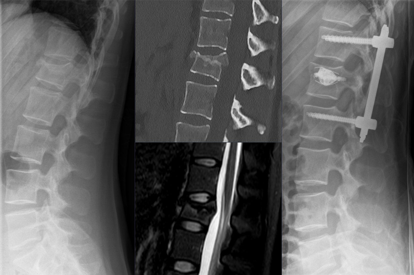
Lumbar Spine
These are common radiograph views of the lumbar spine. Note that the bony prominences of the spine are "flattened" (superimposed) in the AP projection. The junction between the thoracic spine and lumbar spine is seen by identifying the 12th ribs.
Identify the following structures:
- T12 Ribs
- Vertebral Body L1
- Transverse Process L2
- Spinous Process L3
- Pedicle L3
- Inferior Articular Facet L2
- Superior Articular Facet L3
- Intervertebral Disc L3-4
- Intervertebral Foramen L2-3

Identify the following structures:
- T12 Ribs
- Vertebral Body L1
- Transverse Process L2
- Spinous Process L3
- Pedicle L3
- Inferior Articular Facet L2
- Superior Articular Facet L3
- Intervertebral Disc L3-4
- Intervertebral Foramen L3-4

T1-weighted Sagittal MRI
This is a T1-weighted MRI image. Structures containing fat appear bright, while cerebrospinal fluid (CSF), cortical bone and ligamentous structures appear dark.
Identify the following structures:
- L5 Vertebral Body
- L2 Spinous Process
- Spinal Cord
- L3-l4 Intervertebral Disc
- Posterior Epidural Fat Between L3-l4

T2-weighted Sagittal MRI
This is a T2-weighted MRI image. Note that fluid in this type of image, such as the CSF, appears bright. Note the sharp bend (almost 90 degrees) at the junction between the L5 vertebra and sacrum.
Identify the following structures:
- L5 Vertebral Body
- L2 Spinous Process
- Conus Medullaris
- L3-l4 Intervertebral Disc
- Posterior Epidural Fat Between L3-l4

MRI: Axial Lumbar Spine
This image is at a more caudal level than the previous image. The spinal cord is no longer present. What is found in the dural sac inferior to the conus medullaris?
Identify the following
Identify the following structures:
- Vertebral Body
- Spinous Process
- Csf In Subarachnoid Space
- Rootlets Of Cauda Equina
- Dorsal Root Ganglia

Quiz
Radiology
Quiz

Anatomy
Quiz
An abnormal increase in the thoracic curvature of the vertebral column is called:
scoliosis
spina bifida
lordosis
spondylolysis
kyphosis
