
System
Cardiovascular Imaging
Cardiac Imaging Modalities
Radiography
PA Chest Radiography is a quick and low cost means of assessing overall heart size, and may show individual chamber enlargement if it results in a change in the heart shape or contour. The internal anatomy of the heart is not visible on radiography. Chest Radiographs are the best exam to assess for heart failure; specifically for pulmonary edema.
Computed Tomography (CT)
The cardiac chambers, muscles, coronary arteries, large vessels and the pericardium are easily visualized on standard CT scans performed with iv contrast. Computed tomographic angiography (CTA) is a technique in which the CT is performed during a specific early time point after iv contrast administration, when the arteries are brightly enhanced with contrast, but not enough time has passed to allow the contrast to flow through the capillaries and into the veins. CTA is an excellent method to evaluate the coronary arteries, given its high spatial resolution.
CTA datasets can be reconstructed to show the origin and course of the coronary arteries (as well as other, larger arteries) and 3D images of the vessels can be created to show their course and caliber. Since the heart is beating while the CT data is acquired, imaging is performed during both systole and diastole. The CT data can be sampled at specific time points in the cardiac cycle, which enables viewing of the myocardium, papillary muscles and valves during systole and/or diastole, and enables calculation of the left ventricular ejection fraction.
Magnetic Resonance Imaging (MRI)
Magnetic resonance imaging (MRI) has evolved rapidly. MR images are generated based on the magnetic properties of hydrogen ions and basically detect differences in magnetic behavior between hydrogen ions bound in various molecular environments. An MRI examination is made up of multiple imaging series of the heart, each one slightly different. These series are called pulse sequences (which is a standardized way of obtaining an image).
Cardiac MRI/MRA has evolved to become an important tool in the assessment of the heart, and has specific uses in clinical care. You will learn more about this during your clinical rotations & further discussion is beyond the intent of this course.
Angiography
Angiography is a minimally invasive procedure with inherent risks, including arterial trauma and embolization, that although rare, may have serious consequences. Non-coronary angiography is performed by Interventional Radiologists, as well as by other specialties. Coronary Angiography is performed by Interventional Cardiologists, and will be addressed in this course during the cardiology sessions.
Vasculature Imaging Modalities
Radiography
Several blood vessels in the mediastinum are bordered by the lungs, with the contrast of their densities rendering their edges visible. So the edges of the superior vena cava and descending aorta are visible on a radiograph. However, the caliber and luminal patency of the vessels are not visible. Most blood vessels are not conspicuous on radiographs.
Computed Tomographic Angiography (CTA)
Computed tomographic angiography (CTA) is a technique in which the CT is performed during a specific time point after iv contrast administration, when the arteries are brightly enhanced with contrast, but not enough time has passed to allow the contrast to flow through the capillaries and into the veins. CTA is an excellent method to evaluate the vasculature given its high spatial resolution. CTA can be timed to evaluate either the arteries, veins, or both.
CTA datasets can be reconstructed to show the origin and course of arteries throughout the body, and 3D images of the vessels can be created to show their overall size and interrelationships. CTA is widely used to look for aneurysms, to assess for vascular narrowing (stenosis) or occlusion, and to look for acute pathology, like aortic dissection. One major benefit of CTA over angiography is that it demonstrates the arterial lumen, arterial wall and adjacent tissues. Conventional angiography shows ONLY the lumen.
Magnetic Resonance Angiography (MRA)
Magnetic resonance angiography is an alternative method used to evaluate the major arteries and veins, with minimal risk to the patient, and no radiation exposure. It is more expensive and time consuming than CTA or conventional angiography, however it effectively shows the lumen of the large and medium size blood vessels, and is predominantly used to determine vascular patency. Since MRI has lower spatial resolution than CT, it is not as useful for small vessels.
Conventional Angiography
Angiography is a minimally invasive procedure with inherent risks, including arterial trauma and embolization that although rare, may have serious consequences. Angiograms are basically X-ray images of the blood vessels after contrast has been injected directly into them via an intravascular catheter. The contrast opacifies the vessel lumen, rendering it radiodense. It’s important to understand that the images show only the lumen of the blood vessel; the walls and surrounding tissues are not depicted.
Because angiography is invasive, it is no longer considered a first line method of vascular assessment. Instead, it is now used primarily for therapeutic purposes, such as opening occluded or narrowed blood vessels, stopping bleeding, and treating other acute conditions.
Ultrasound
Ultrasound is a relatively low cost and low risk means of evaluating the heart (called echocardiography- performed by cardiologists) and blood vessels, and is used extensively in the carotid arteries. Doppler ultrasound informs about the overall caliber of a vessel, provides information about the vessel wall and lumen. It effectively demonstrates stenosis (narrowing). Spectral doppler is used to measure flow velocity to help determine if an area of narrowing is significantly limiting blood flow.
Heart Structures
PA Chest Radiograph
The size of the heart and the borders of certain chambers can be evaluated on a PA chest film. By performing the film PA, the heart is placed close to the detector plate, preventing magnification of the heart.
Identify the following structures:
- Right Heart Border
- Left Heart Border
- Aortic Arch
- Superior Vena Cava
- Right Atrium
- Left Ventricle
- Apex

Cardiomegaly
This is a comparison of a normal-sized heart (right image) with an enlarged heart (left image). Notice that the enlarged heart is more globular in shape. The left heart borders is closer to the left lateral chest wall, and the right heart border extends out farther from the spine compared to the normal heart.

Lateral Chest Radiograph
In this view, the anterior and posterior heart borders are visible. There is some superimposition of the heart chambers on this view. The most posterior chamber is the left atrium, and the most anterior chamber is the right ventricle.
Identify the following structures:
- Right Ventricle Border
- Left Atrium Border
- Aortic Arch

CT Heart
This is an axial CT image of the heart. Below are a reference PA chest radiograph and a comparable anatomic image from the Visible man showing the approximate level of this axial slice. As in all axial (transverse) images, the patient is lying supine and you are observing from the patient's feet looking towards the head.
An iodine based CT contrast agent was injected intravenously to opacify (increase the density of) the heart chambers and blood vessels.
Identify the following structures:
- Sternum
- Left Ribs
- Right Pulmonary Vein
- Left Atrium
- Left Ventricle
- Mitral Valve
- Right Atrium
- Right Ventricle
- Interventricular Septum
- Descending Aorta


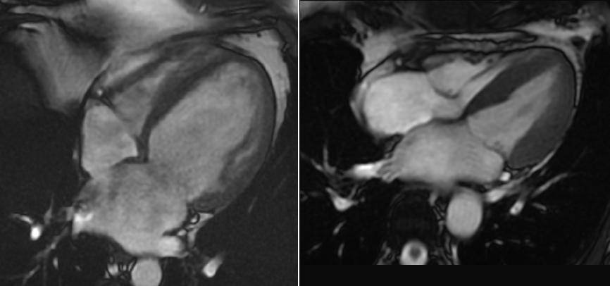
Coronal CT
This image is of the same patient as in the previous image. CT images are obtained as a helix of data that can then be reformatted into imaging planes. This is a coronal reformated image of the contrast enhanced chest CT.
Identify the following structures:
- Left Venticle
- Myocardium
- Ascending Aorta
- Aortic Valve
- Pulmonary Trunk
- Right Atrium
- Superior Vena Cava
- Interventricular Septum
- Right Ventricle
- Diaphragm

Cardiac Chambers
The borders of each cardiac chamber are not discernable on radiography (except where they are border forming with the lung, like the lateral margin of the right atrium). This demonstrates the approximate position of the chambers and large vessels.
Identify the following structures:
- Right Ventricle
- Left Ventricle
- Right Atrium
- Pulmonary Arteries
- Superior Vena Cava
- Ascending Aorta

Cardiac Chambers on Radiography (Lateral)
This demonstrates the approximate position of the chambers and large vessels on the lateral radiograph.
Identify the following structures:
- Right Ventricle
- Left Ventricle
- Left Atrium
- Main Pulmonary Artery

Mediastinum
PA Chest Radiograph
Many of the mediastinal structures are of similar density, so it is difficult to differentiate them with radiography. However, structures that are adjacent to the radiolucent lungs (less dense) can be visualized due to the difference in density.
Identify the following structures:
- Heart
- Aortic Knob
- Superior Vena Cava
- Descending Aorta
- Trachea
- Trachial Bifurcation

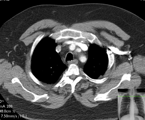
CT: Axial Superior Mediastinum
This is a series of axial (transverse) CT images of the thorax of one patient. The images have been "windowed" (contrast adjusted) in order to highlight soft-tissue structures, and consequently, the lungs appear mostly black. As you work through these images, it may be helpful to remember the structures in the previous "slice" in order to be able to trace those structures and maintain orientation. This first image will ask you to name several structures for reference, most of which will continue to be present in subsequent slices. In all of these images, vascular structures are bright due to the injection of contrast dye in the venous system (usually via the arm) of the patient. You can assume that black represents air. The reference chest radiographs show the approximate level of the axial slice.
Slices
Each of the following CT slices is more caudal than the slice prior.
Identify the following structures:
- Manubrium
- Vertebral Body
- Spinal Cord
- Spinous Process
- Ribs
- Pectoralis Major
- Subcutaneous Adipose Tissue
- Scapulae
- Trachea
- Esophagus
- Left Brachiocephalic Vein
- Right Brachiocephalic Vein
- Brachiocephalic (Innominate) Artery
- Left Common Carotid Artery
- Left Subclavian Artery
Vasculature
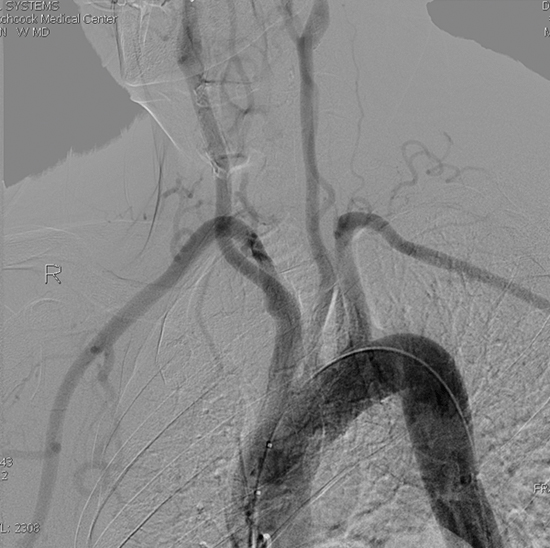
Aortic Arch Angiogram
An aortic arch angiogram is obtained by threading a catheter into the ascending aorta and injecting iodine-based dye. Sequential X-rays are then taken as the dye is injected. This image is a static image from the angiogram series.
Identify the following structures:
- Ascending Aorta
- Brachiocephalic Artery
- Left Common Carotid Artery
- Left Subclavian Artery
- Left Vertebral Artery
- Right Common Carotid Artery
- Right Subclavian Artery
- Right Internal Thoracic Artery
Left Coronoary Angiogram

An angiogram of the coronary arteries is performed by threading a small catheter from the femoral artery in the groin up into the aorta, and selectively cannulating the coronary ostia. Radio-opaque dye is then injected and X-ray images are obtained via fluoroscopy to evaluate the coronary anatomy. Fluoroscopy allows for real-time acquisition of images, and is a technique which is conceptually similar to the standard X-ray radiographs which you have seen thus far. A major difference is that radio-dense material appears dark rather than bright. This image of the left coronary system is obtained via a right anterior oblique view of the heart.
Identify the following structures:
- Anterior Interventricular Artery (Lad)
- Anterior Interventricular Artery (Lad)
- Left Coronary Artery (Left Main Artery)
- Circumflex Artery
- Obtuse Marginal Branch Of The Circumflex Artery
- Atrioventricular Groove Branch Of The Circumflex Artery

Right Coronary Angiogram
This image was obtained via a left anterior oblique view (almost as thought you were looking "down the barrel" of the heart) in order to view the right coronary artery as it courses along the outside of the atrioventricular groove on the right. Because of the angle, the posterior interventricular artery branch will look significantly foreshortened.
Identify the following structures:
- Angiogram Catheter
- Right Coronary Artery
- Branch To Sinoatrial Node
- Posterior Interventricular Artery

Left Ventriculogram
This image is obtained by threading the catheter into the left ventricle and injecting contrast dye. The dynamic ejection of blood into the aorta can then be evaluated.
Identify the following structures:
- Angiogram Catheter
- Left Ventricle (Filling With Contrast Dye)
- Aorta
- Aortic Valve

Imaging the Heart Video
Linked below is a 10 minute introduction into imaging the heart recorded by Nancy McNulty, MD (Professor of Radioloy and Medical Education) that is intented to supplement the material covered in class.
Questions
Anatomy Quiz
Quiz
Which structure is associated with the right ventricle?
Coronary sinus
Fossa ovalis
Mitral valve
Moderator band (septomarginal trabecula)
Pectinate muscles
Practical Quiz
Quiz
Identify the labeled structure.

Superior vena cava
Right atrium
Right ventricle
Aorta
Right coronary artery
Radiology Quiz
Quiz
Identify the labeled chamber of the heart.

Right atrium
Right ventricle
Left atrium
Left ventricle