
System
Pulmonary Imaging

PA and Lateral Chest Radiographs
A chest X-ray (CXR) is one of the most common imaging tests you will encounter as a clinician. Two views (PA and lateral) are obtained at 90 degrees from each other in order to gain an appreciation of dimensionality.
Structures of the chest wall can be evaluated on radiographs. The breast tissue cannot be evaluated on standard CXR due to poor visualization of the breast parenchyma.


Chest Wall
Lateral Chest
In the lateral view, the anterior and posterior limits of the thoracic cavity are visible.
Identify the following structures:
- Sternum
- Vertebral Column
- Breast

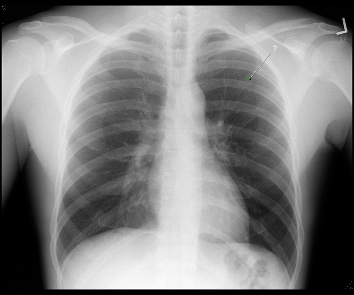
PA Chest
This is a normal PA chest xray. When the image is taken, the scapula is protracted to minimize its overlap with the chest wall. Can you see where the medial border of the scapula is overlapping with the chest wall? Notice that the anterior and posterior parts of the ribs are superimposed. The portion of the rib that is more horizontal is the posterior part, while the anterior part of the rib slopes from superior to inferior.
Identify the following structures:
- Clavicle
- Scapula
- Ribs

BONUS: Counting ribs on a CXR is a skill that requires some practice. Radiologists typically find rib 1 and count from there. On the right side of the patient, see if you can find the intersection between the anterior part of the 5th rib and the posterior part of the 8th rib. When you add the labels to the image a dot will appear at this intersection.
Lung Anatomy
PA Chest
Air does not absorb X-ray beams, so areas filled with air, such as the lungs and bronchial tree, appear dark on a CXR. Notice that there are white "streaks" in the lungs, especially near the hilum. These streaks are formed by the branches of the bronchial tree and pulmonary vessels. Notice that in a normal chest film, the right hemidiaphragm is slightly higher than the left hemidiaphragm.
Identify the following structures:
- Trachea
- Right Lung
- Left Lung
- Right Hemidiaphragm
- Left Hemidiaphragm

Lateral Chest
A lateral film is taken with the left side of the patient against the detector plate to reduce magnification of the heart (structures farther from the detector become magnified). Because of this, the right ribs are slightly magnified and appear larger than the left ribs. Notice that the right and left lungs are superimposed on a lateral film.
Identify the following structures:
- Left Lung
- Right Lung

Lobar Anatomy - PA
Portions of each lobe of the lung are superimposed on one another on a CXR. Because of this, it is not always possible to pinpoint the lobar location of a nodule on a PA CXR.
Identify the following structures:
- Left Upper Lobe
- Left Lower Lobe
- Right Middle Lobe

Lobar Anatomy - Lateral
The lower lobes of each lung are completely superimposed on one another on a lateral CXR, and portions of the upper and right middle lobes are superimposed. Because of this, it is not possible to determine which lung an abnormality is in on a lateral CXR.
Identify the following structures:
- Left Upper Lobe
- Left Lower Lobe

Pulmonary Vasculature
Pulmonary Angiogram
This image is obtained by threading a thin catheter into the pulmonary vasculature and injecting iodine-based dye. Iodine is radio-dense and relatively well tolerated. An X-ray image is then taken immediately in order to visualize the arterial flow pattern.
Identify the following structures:
- Angiogram Catheter
- Left Pulmonary Artery
- Right Pulmonary Artery
- Right Upper Lobe Trunk
- Right Middle Lobe Artery
- Right Lower Lobe Trunk
- Left Upper Lobe Branches
- Left Lower Lobe Banches

Venogram

Compare the arteriogram above with the pulmonary venogram below obtained during the venous phase of the blood flow.
Questions
Radiology Quiz
Quiz
Which part of the rib is labeled?
anterior
posterior
Anatomy Quiz
Quiz
The horizontal fissure separates:
left upper lobe from left middle lobe
left middle lobe from left lower lobe
left upper lobe from left lower lobe
right upper lobe from right middle lobe
right middle lobe from right lower lobe
right upper lobe from right lower lobe
